
Researchers develop nanoparticles from viruses to deliver drugs in our body
Aditi Karmarkar Bengaluru Jan 30,
Viruses are often looked down with disdain as they cause many diseases—from the seasonal flu to the incurable AIDS. However, not all viruses are harmful, and some help us solve some of the biotechnological problems like drug delivery for the treatment of various diseases. Most often, these are plant viruses, which are not infectious to us, are biodegradable and can be produced on a large scale. In a recent study, researchers from the Indian Institute of Science (IISc), Bengaluru, and Sri Venkateswara University, Tirupati, have developed nanoparticles that can deliver drugs to targeted mammalian cells, using a type of plant virus called sesbania mosaic virus.
Viruses have a protein covering called capsid, inside which the genetic information lies in the form of RNA or DNA. Due to their small size, viruses can be modified to act as nanoparticles that carry drugs or dyes, thus having therapeutic and imaging applications. In this study, published in the journal Archives of Virology, the researchers have added fluorescent tags to the sesbania mosaic virus to help in imaging. The research was supported by the Department of Biotechnology (DBT), Department of Science and Technology (DST) and the Indian National Science Academy (INSA).
The sesbania mosaic virus affects the Hummingbird tree (Sesbania grandiflora). In a previous study, the researchers have shown that this virus was a good candidate for creating nanoparticles as it could tolerate the addition of amino acids, which are present in many drugs and dyes, to the capsid without affecting its structure. In this study, they have developed such viral nanoparticles by identifying suitable sites on the capsid that could be modified. By studying the crystal structure of capsid, they found that the amino acids present on its external and internal surface can be used to bind other molecules of interest.
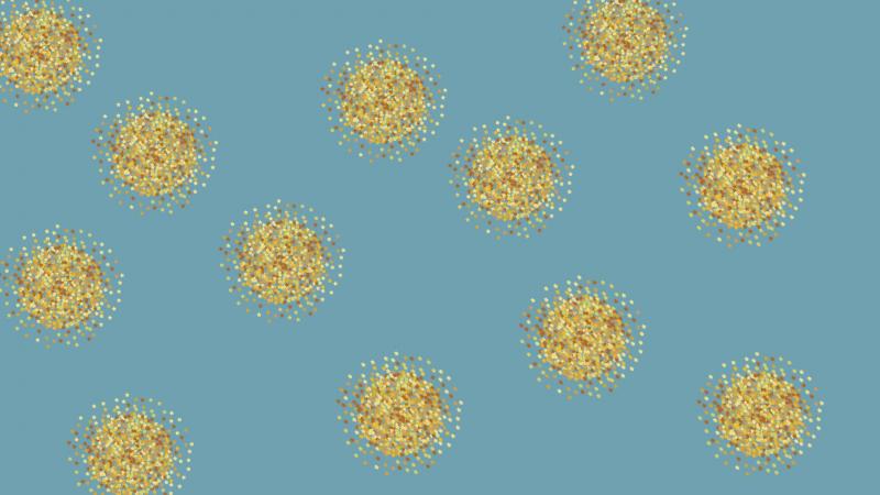
The researchers successfully bound the fluorescent dye Alexa Fluor 488 to the external surface of the capsid and the dye Cyanine 5.5 maleimide to the internal surface. The viral nanoparticles thus formed were stable and assembled into a proper icosahedral structure—a geometric structure with 20 faces. When the two types of nanoparticles with different dyes were tested on animal cells, the researchers found that the nanoparticles with Cyanine 5.5 maleimide showed the highest fluorescence intensity for imaging applications.
So, how do these nanoparticles interact with our cells? The researchers found that the viral nanoparticles started by first interacting with surface proteins of animal cells, like vimentin, to enter the cell. Vimentin is a cytoskeletal protein that controls the internal architecture of the cell. This interaction probably results in the cell engulfing the nanoparticle, but other modes of internalisation cannot be ruled out yet.
The nanoparticles developed using sesbania mosaic virus are non-toxic, biocompatible and easy to produce on a commercial scale. The researchers believe that in the future, these particles could be modified and incorporated into human cells for diagnosing and treating many diseases.
“These particles were shown to have a natural preference for certain cells, which could be exploited further for targeted delivery of drugs and for in vivo imaging for future use in cancer diagnosis and therapy,” the scientists say.
Perhaps viruses are now turning out to be heroes more than the villains that they once were thought to be!
Image and news Source: Development of sesbania mosaic virus nanoparticles for imaging, Research Matters